


After thoracotomy, pulmonary resection, and mediastinal lymph node dissection, a patient is determined to have a squamous cell carcinoma 2 cm. in diameter, located 1 cm. from the carina along the right mainstem bronchus. Three peribronchial lymph
belongs to book: ASIR SURGICAL MCQs BANK|Dr. Gharama Al-Shehri|1st edition| Chapter number:11| Question number:35
All Answers
total answers (1)
 Medicine
C. T2N1M0 Stage II.
DISCUSSION: The TNM staging system for carcinoma of the lung provides a consistent, reproducible description of the anatomic extent of disease at the time of diagnosis. In the TNM system, T represents the primary tumor and numerical suffixes describe increasing size or involvement; N represents regional lymph nodes with suffixes to describe levels of involvement; and M designates the presence or absence of distant metastases.
TUMOR (T)
TX Occult carcinoma (malignant cells in sputum or bronchial washings but tumor not visualized by imaging studies or bronchoscopy)
T1 Tumor 3 cm. or less in greatest diameter, surrounded by lung or visceral pleura, but not proximal to a lobar bronchus
T2 Tumor larger than 3 cm. in diameter, or with involvement of main bronchus at least 2 cm. distal to carina, or with visceral pleural invasion, or with associated atelectasis or obstructive pneumonitis extending to the hilar region but not involving the entire lung
T3 Tumor invading chest wall, diaphragm, mediastinal pleura, or parietal pericardium; or tumor in main bronchus within 2 cm. of, but not invading, carina; or atelectasis of obstructive pneumonitis of the entire lung
T4 Tumor invading mediastinum, heart, great vessels, trachea, esophagus, vertebral body, or carina; or ipsilateral malignant pleural effusion
NODES (N)
N0 No regional lymph node metastases
N1 Metastases to ipsilateral peribronchial or hilar nodes
N2 Metastases to ipsilateral mediastinal or subcarinal nodes
N3 Metastases to contralateral mediastinal or hilar, or to any scalene or supraclavicular nodes
DISTANT METASTASES (M)
M0 No distant metastases
M1 Distant metastases
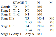
The TNM subsets are subsequently grouped in a series of stages of disease to identify groups of patients with similar prognosis and therapy.
need an explanation for this answer? contact us directly to get an explanation for this answer