A 42-year-old woman is subjected to computed tomography (CT) scan of the abdomen for recurrent complaints of epigastric discomfort which revealed a 2.6 cm × 2 cm × 3 cm well rounded mass in her left adrenal gland, with a attenuation value of 5 HU, well enhancing on contrast administration. Washout study is not done. She was diagnosed with hypertension and diabetes since 2 years for which she is on amlodipine 5 mg/day and metformin 2 g/day. She has progressively gained weight since the birth of her last child birth ten years ago and currently is obese with a BMI 28 kg/m2. Her menstrual cycles are irregular since 5 years and bleeds every 2 months only after ingestion of a pill taken twice daily for a week. On examination, central obesity is present, supraclavicular areas are full. Skin appears normal with no thinning, bruising or striae. No proximal myopathy or hirsutism is noted.
Management: The imaging characteristics indicate a benign lesion. Her clinical profile validates screening for pheochromocytoma, Cushing’s syndrome and primary hyperaldosteronism. All her tests are negative. She is advised to follow-up every 6 months with repeat imaging and biochemical testing.
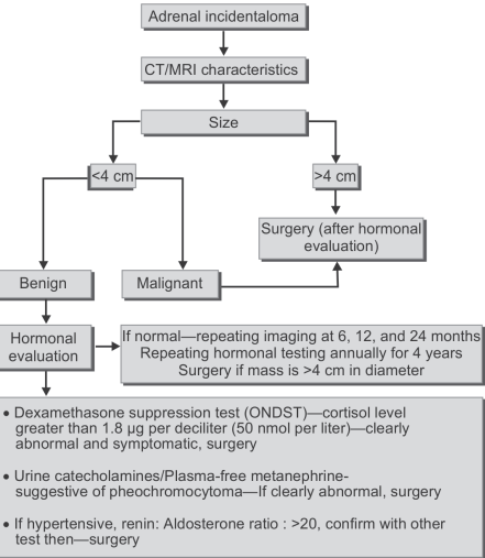
What is the management of adrenal incidentaloma?


 Medicine
need an explanation for this answer? contact us directly to get an explanation for this answer