


0
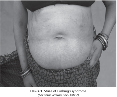
A 29-year-old female presented with progressive swelling over the body, weight gain, menstrual irregularity. On examination, her BP was 160/100 mm Hg, pulse rate 86/min, weight 88 kg, height 155 cm. Buffalo hump was present with broad purplish striae over the abdomen. Proximal muscle weakness was present. Her investigations revealed Hb 12.6 g%, fasting blood sugar (FBS) 170 mg%, creatinine 0.9, Na 140 mEq/L, K 3.4 mEq/L (Fig. 2.1).
What imaging would be required in Cushing’s syndrome?
 Medicine
Once the diagnosis of ACTH or non-ACTH-dependent Cushing’s syndrome is made appropriate imaging is done. In non-ACTH-dependent Cushing’s CT/MRI of the adrenal gland is done but CT gives the better resolution of the adrenal anatomy. In ACTH-dependent Cushing’s syndrome, the first step is to image the pituitary, and see if there is any abnormality and for this MRI of the pituitary is done with gadolinium enhancement. Use of dynamic MRI (with IV gadolinium) sequences increases the sensitivity of detection. However, caution has to be exercised in diagnosing microadenomas as the possibility of an incidentaloma is as high as 10% in general population. If the pituitary imaging is negative, then imaging of the thorax, abdomen head and neck has to be done to detect the ectopic source of production of the ACTH [mostly thymoma, carcinoid, pheochromocytoma, medullary thyroid carcinoma (MTC) or malignancy]. In patients with suspected ectopic ACTH and not localized, special imaging like 18F-fluorodeoxyglucose positron emission tomography (FDG-PET) may be done. Inferior petrosal sinus sampling (IPSS) is an invasive procedure that involves measuring the central-to-peripheral ACTH gradient and may be useful when the source of ACTH production (i.e. pituitary versus ectopic) remains elusive despite other noninvasive testing [i.e. when there is cortisol suppression on HDDST (suggesting Cushing’s disease) in the setting of a negative pituitary MRI].
need an explanation for this answer? contact us directly to get an explanation for this answer