


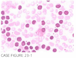
A 62 -year-old male presen ts for evaluation of bilateral cervical, axi l lary, and inguinal lymphadenopathy. Physical exami nation con firms these fi ndi ngs in addition to disclos-ing splenomegaly. Laboratory evaluation reveals leukocyte count of 70,000/pL hemoglobin of 9.6 g/dl, and platelet count of 1 38 , 000/JLL. Flow cytometry reveals increased population of cel ls expressing CDS , CD 1 9 , CD2 0, and CD2 3. Peripheral blood smear is shown below.
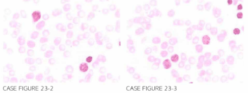
Patien t was treated wi th 6 cycles of bendamustine and rituximab with normal ization of peripheral blood coun ts and disappearance of lymphadenopathy. Over the next 3 years, the patien t was fol lowed with no evidence of active disease. However, during the fourth year of follow-up, he presen ted wi th multiple new skin lesions and fever. Labo-ratory fi ndi ngs revealed leukocyte count of 27,000/JLL, hemoglobin of 1 1 .9 g/dl, platelet cou nt of 1 2 Lj,OOO/JLL, and LDH of 5Lj7 U/L. Peripheral blood smear is shown
What is the most likely explanation of his presentation?
- Transformation to prolymphocytic leukemia (PLL)
- Transformation to aggressive lymphoma
- Transformation to acute lymphoblastic leukemia (ALL)
- Therapy-related myelodysplastic syndrome (t-MDS)
- Relapse of chronic lymphocytic leukemia (CLL)
 Medicine
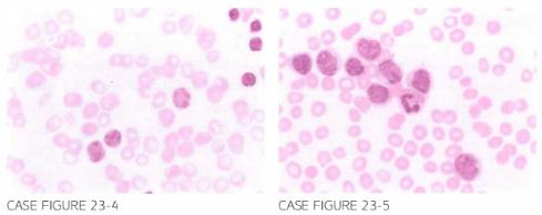
B. Peripheral blood slide reveals a different cell population from that observed originally at diagnosis. The abnormal cells are large with sparse cytoplasm, fine uncondensed chromatin, and no nucleoli. These cells do not resemble prolymphocytes that are characterized by their large size, some degree of chromatin condensation, increased amount of cytoplasm, and prominent nucleolus. Taken together with new skin lesions, B symptoms, and elevated LDH, the presentation is most consistent with a transformation to an aggressive lymphoma, Richter's transformation (RT). Skin biopsy was ob-tained and histiologic exam revealed diffuse large B-cell lymphoma (DLBCL). DLBCL is the most common histology seen in RT; however, rare cases of Hodgkin lymphoma and even T-cell lymphomas have been reported. Below is a comparison between the peripheral blood picture in RT and PLL (Cancer. 2005;103(2):2 1 6, Br J Haematol. 20 12;156(1):50, Am J Clin Pathol. 1 990;93(3):333, Am J Clin Pathol. 1995;103(3):348).
need an explanation for this answer? contact us directly to get an explanation for this answer