


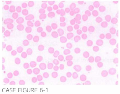
A 32 -year-old female is found on routine physical examination to have splenomegaly. She reports no acu te change in her energy levels. However. she does note bei ng more easily fatigued duri ng heavy physical activity as long as she can remember. Patien t re-ports a h istory of cholelithiasis diagnosed 1 0 years previously; however. she elected not to undergo cholecystectomy. Mild splenomegaly is noted on physical examination . Laboratory workup reveals hemoglobin 11 .2 g/dl. mean corpuscular volume (MCV) 85 fl. leu kocyte count 6.500/,LLL. and platelet count of 361 . 000/,LLL. I ndirect bil irubin level ismini mall el vated and tneoireccanriglooin--resns negative. -reriph-eraJ-blood· smear is shown below.
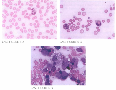
Patient underwent splenectomy with normal ization in her hemoglobin level. Two years after the procedure. she presen ts to the emergency departmen t with acute onset of fever. con fusion . and hypotension refractory to aggressive intravenous fluid admin is- tration. Laboratory evaluation reveals pancytopenia with acute rena� fai lure. Blood cul-tures returns posi tive for Gram-negative bacteria as shown below. Peripheral blood smear and bone marrow aspi rate are reviewed and are shown below.
What is the most likely explanation for her clinical presentation?
- Autoimmune hemolytic anemia with immune thrombocytopenia
- Gram-negative sepsis with multiorgan failure
- Hemophagocytic syndrome
- Acute myeloid leukemia
- Thrombotic thrombocytopenic purpura
 Medicine
C. Peripheral blood smear reveals erythrophagocytosis, and bone marrow aspirate shows hemophagocytosis. This blood smear picture, in addition to her presentation with pancytopenia and Gram-negative sepsis in a postsplenectomy setting, is most consistent with secondary hemophagocytic syndrome associated with bacterial infection. Answer B is partially correct, however, as it does not explain the peripheral blood picture and bone marrow images (Cancer. 1984;54(12):2968).
need an explanation for this answer? contact us directly to get an explanation for this answer