


History
A 4-year-old boy is referred from the accident and emergency department to the paedi-atric dermatology clinic, owing to a 1-year history of scaling and crusting of his scalp associated with hair loss. Despite numerous courses of oral antibiotics and topical anti-fungal creams the eruption had become progressively worse over the last few months. No one else at home is obviously affected. The child is otherwise well.
Examination
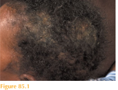
The patient is accompanied by his parents and older sibling. There are obvious patches of alopecia over the vertex of his scalp associated with scaling and crusting (Fig. 85.1). The skin is inflamed and erythematous in the affected areas. There is marked occipital lymphaden- opathy. The rest of his skin and nails look nor-mal. Examination of his older sibling reveals a diffusely scaly scalp, but no alopecia or lym-phadenopathy. The patient’s mother is noted to have a scaly annular lesion on her anterior neck, both parents’ scalps look normal.
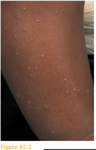
The patient was started on oral therapy daily plus a shampoo, but within five days he had developed a widespread itchy papular rash (Fig. 85.2) and returned to the dermatology clinic.
Questions
• What disease is affecting the patient’s scalp and what is the cause of his wide-spread rash?
• Which members of the family would you treat and with what agents?
• Is the patient’s hair likely to grow back?
 Medicine
This boy presents with a very common scalp complaint of children seen in most urban areas. He is suffering from tinea capitis (scalp ‘ringworm’) which is a dermatophyte fungal infection. The clinical appearances can be highly variable from marked scaling, crusting,
pustules and papules to a frank inflammatory boggy swelling (kerion) with alopecia, to sub-tle fine diffuse scale only. Children frequently have associated occipital lymphadenopathy.
After starting oral antifungal medication the patient developed a classic pruritic papular ‘id’ reaction which is an immunological reaction to the infection that can coincide with the start of treatment. It is not an allergy to the antifungal treatment as is frequently assumed. The ‘id’ reaction can be managed with mild topical steroids and emollients. The oral antifungals should be continued. The fungus is spread from child to child in schools and between family members at home.
Frequently, parents’ scalps are spared, however they may develop cutaneous lesions (tineacorporis), especially on their neck and upper trunk where the child’s head comes to rest whilst sitting on the parent’s lap.
Trichophyton tonsurans is an anthropophilic (human) species, which is most commonly isolated from the mycological culture. The fungal hyphae of T. tonsurans penetrate intothe hair shaft (endothrix) rendering topical therapy ineffective. Therefore, systemic treat-ment is required to clear the scalp infection. Screening of family members by taking scalp brushings is recommended as tinea capitis may be very subtle clinically. Mycology cultures can take 6–8 weeks, as the fungus grows slowly; consequently, treatment may need to be started in the first instance on clinical grounds.
Traditionally, children with tinea capitis were treated with oral griseofulvin, however to eradicate T. tonsurans high doses are required for up to two months. Oral terbinafine is therefore the treatment of choice in many paediatric dermatology clinics, as it is highly effective and well tolerated. As yet, oral terbinafine is not licensed in children in the UK. Oral terbinafine is given to children daily for 1 month according to their weight: < 20 kg, 62.5 mg; 20–40 kg, 125 mg; > 40 kg, 250 mg. It is good clinical practice to re-brush the scalp after treatment to ensure that there is a mycological as well as clinical cure.
In this case the two children should be treated with oral antifungal medicine and their mother given topical terbinafine 1% cream to treat her tinea corporis. It is a good idea to ask all the family members to use an antifungal shampoo once/twice weekly to reduce the spread of fungus during the treatment course. Fortunately, the alopecia has a very high chance of complete recovery after treatment, even after a highly inflammatory kerion.
KEY POINTS
• Clinical features of tinea capitis are highly variable, from mild diffuse scale to pustules and swelling.
• Affected children and their close contacts should be screened for tinea capitis by scalp brushings.
• Systemic antifungals should be given, as the majority of cases are caused by endothrix fungi.
need an explanation for this answer? contact us directly to get an explanation for this answer