


History
A 5-year-old boy presents to the accident and emergency department with a sudden-onset facial eruption over the previous 24 hours. Initially the skin was erythematous and crusted, then his parents noticed blisters developing and were understandably concerned.
Apart from a fever the previous day he is otherwise well, and is eating and drinking nor-mally. He is recovering from a recent episode of chickenpox but otherwise has had no previous skin problems. No one else at home is obviously affected.
Examination
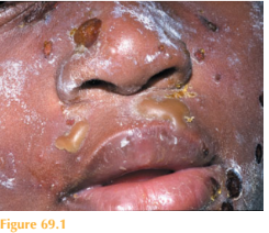
There are crusted resolving chickenpox lesions on his face, trunk and limbs. On his cen-
tral face there are acute tense bullae and areas of golden crusting (Fig. 69.1), but no ulcers or erosions. Calamine lotion applied to the skin by his parents has given him a patchy white appearance. Elsewhere on the trunk and limbs there are healing crusted lesions and post-inflammatory hyperpigmentation. He is now apyrexial and has normal blood pressure and pulse rate.

Questions
• Why do blisters form?
• What is the differential diagnosis?
• Who else may be affected?
 Medicine
Blisters (vesicles and bullae) are usually a sign of an acute cutaneous reaction. Blisters comprise clear, serous fluid, which may become cloudy due to the accumulation of neu-trophils (pus), or blood-stained due to vascular damage. Blisters can form due to damage to the adhesion structures in between cells; this damage can result from friction/pressure,heat, infections, autoimmune-mediated complexes, phototoxic reactions, adverse drug reactions, and congenital deficiencies or malfunction of adhesion structures within the skin. Blisters can also form due to cutaneous oedema. This child has bullous impetigo, which results from a cutaneous infection with exotoxin-producing strains of Staphylococcus aureus (confirmed by skin swabs). Exotoxins cause loss of cell–cell adhesion in the epidermis, resulting in superficial blisters which break down, leaving denuded areas and classic golden crusting. Impetigo is very common, especially in children, and is highly contagious with spread by direct inoculation. The initial infection may occur through a small break in the skin – this child had resolving chickenpox lesions which left his skin vulnerable to secondary bacterial infection. Other family members are commonly subsequently infected by contact with the index case. Classically, impetigo develops rapidly on localized areas of skin. Blisters, pustules, erosions and golden crusted exudate complete the clinical picture. Patients are usually otherwise well. Skin swabs may isolate S. aureus or occasionally Streptococcus (group A). Localized cutaneous impetigo can be managed with topical antiseptics such as dilute chlorhexidine to wash the skin and topical antibiotics such as fusidic acid, polymyxin or mupirocin. Oral antibiotics such as flucloxacillin or erythromycin may be required if involvement is more widespread.
KEY POINTS
• Blisters indicate an acute cutaneous reaction.
• Impetigo is common and highly contagious.
• Patients with localized disease may be managed with topical antibiotics.
need an explanation for this answer? contact us directly to get an explanation for this answer