


History
A 54-year-old woman presents with a widespread eruption that has persisted for almost 20 years. She had initially noticed the first few patches over her pelvic girdle, but slowly over the years the patches have become more widespread. The patches are occasionally mildly itchy; she has noticed a slight dryness over their surface. She has tried over-the-counter antifungal creams and emollients with no real benefit. She is systemically well and takes hormone replacement therapy.
Examination
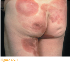
There is an extensive eruption of multiple erythematous patches with overlying scale over the trunk and limbs, occurring predominantly in unexposed areas (Fig. 65.1). The patches are oval to annular in shape and most, but not all, are well demarcated. Some of the areas are thicker, forming thin plaques.

Questions
• What is the diagnosis?
• What are the differential diagnoses?
• What are the options for treatment?
 Medicine
Any patient presenting with a very long history of a slowly progressive skin eruption that is unresponsive to topical therapy warrants a skin biopsy. The diagnosis that shouldn’t be missed is mycosis fungoides, a form of cutaneous T-cell lymphoma (CTCL). This patient is typical of those presenting with an indolent rash over many years that has a predilection for the pelvic girdle. The condition in the early stages classically presents with erythema-tous patches with fine overlying scale.
CTCL encompasses 65 per cent of all cutaneous lymphomas. Mycosis fungoides is the commonest of these, accounting for almost half of all cases. It is a low-grade T-cell lymphoma in which there is an abnormal neoplastic proliferation of lymphocytes of a ́T ́ subtype (thymus-derived). There is a male predominance and it has an incidence of 1 per 100 000 per year.
The average time from disease onset to diagnosis is 7 years. Patients may be misdiagnosed with psoriasis, parapsoriasis, discoid eczema or tinea corporis. Even for an experienced dermatologist the diagnosis may be a challenge, as several skin biopsies may be required to finally clinch the suspected diagnosis of CTCL. Histopathology classically shows atypi-cal lymphocytes in the upper dermis extending into the epidermis with epidermotropism and Pautriers’ microabscesses. T-cell gene rearrangement studies from skin biopsies can help to confirm the diagnosis by showing a clone of abnormal T-lymphocytes. CTCL has three skin stages: 1. Patch stage: macular erythema to patches with overlying scale predominantly over non-exposed skin. Spontaneous resolution, fixed lesions and slow progression may be seen. Involved skin can be pruritic and atrophic (thinned).
2. Plaque stage: The patches become thickened and form plaques.
3. Tumour stage: Large irregular nodules develop within plaques, or de novo. At this stage, spread to other organs is more likely than in the earlier stages. CTCL may remain confined to the skin for many years running an indolent course, but the abnormal cells can eventually infiltrate other tissues including blood, lymph nodes and visceral sites, usually in the context of extensive cutaneous involvement with wide-spread plaques, tumours or erythroderma. Unlike some other lymphomas, the prognosis is generally good. However, acurate diagnosis and staging is critical in determining the prognosis of those with CTCL. Investigations therefore are tailored to the clinical setting. Management of patients should be carried out by a multidisciplinary team. Currently, treatment is not curative but is aimed at controlling symptoms and cosmesis whilst limiting toxicity. Early-stage disease is treated with skin-directed therapy such as topi-cal corticosteroids, phototherapy (psoralen–UVA) and radiotherapy. Patch-stage disease usually improves with topical and phototherapy whereas more infiltrated plaques may require radiotherapy. More advanced disease requires more aggressive treatment includ-ing extracorporeal photophoresis, oral retinoids, interferon, single-agent and multi-agent chemotherapy, and even stem-cell bone marrow transplantation.
KEY POINTS
• Mycosis fungoides is the most common form of cutaneous T-cell lymphoma.
• It is an indolent rash that has a predilection for the pelvic girdle.
• It may infiltrate other tissues including blood, lymph nodes and visceral sites.
need an explanation for this answer? contact us directly to get an explanation for this answer