


History
A 73-year-old man presents with an 18-month history of a pigmented lesion on his left cheek. He had not paid much attention to it and had assumed it to be a ‘sun spot’; however, his daughter is concerned that it has been enlarging slowly and appears to be getting darker in colour. She had taken her father to the GP who had then referred him to the dermatology out-patient department. The patient is a retired roofer and keen gar-dener. He takes aspirin and an antihypertensive medication.
Examination
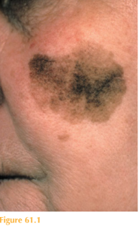
The patient has a brown to black macular lesion measuring 2 cm in diameter that has a highly irregular border over his left cheek (Fig. 61.1). He is tanned but otherwise his full skin check is unremarkable.

Questions
• What is the most likely diagnosis?
• What diagnosis should be excluded?
• How would you manage the patient?
 Medicine
Patients presenting with large pigmented lesions on their skin appearing in later life always warrant referral to a dermatology specialist. This elderly patient had an outdoor occupation before retirement and now spends long hours outside in the garden. Clinically the most likely diagnosis is lentigo maligna (LM). These lesions usually appear in the seventh to eighth decades of life. Sun-exposed sites including the face, neck and dorsi of the hands are most commonly affected. Large macules appear with highly irregular borders and significant variation of pigmentation from tan, brown to black. LM can be a precursor for malignant melanoma in situ, so-called lentigo maligna melanoma (LMM). Dermatologists have a low threshold for performing a biopsy from these lesions to exclude malignant transformation. Sampling such large lesions with small punch biopsies, however, may lead to sampling errors and the clinical appearance of the lesion is also important in determining the ultimate management.
Histopathologically, LM is characterized by abnormal melanocytes confined within the epidermis above the basement membrane. When atypical melanocytes invade the rich vascular and lymphatic networks of the dermis, then this becomes LMM.
Possible risk factors for LM include ultraviolet light exposure, fair skin types and occu-pational risks. Differential diagnoses include solar and seborrhoeic keratoses and LMM.
The definitive treatment of LM is surgical excision. In many patients, however, surgery may not be possible due to other co-morbidities. Other modalities of treatment that may be considered include radiotherapy, cryotherapy and topical imiquimod 5% cream (which is an immunomodulator).
The development of a papular or nodular element within the LM should lead to a high suspicion of transformation to melanoma. LMM makes up 15 per cent of UK malignant melanomas.
KEY POINTS
• Lentigo maligna (LM) occurs in the seventh or eighth decades on sun-exposed skin.
• LM can be a precursor for malignant melanoma.
• The definitive treatment of LM is surgical excision.
need an explanation for this answer? contact us directly to get an explanation for this answer