


0
History
A 28-year-old woman presents with multiple naevi, which had gradually appeared over many years, since puberty. She denies any history of obvious change in her moles but feels she has had too many to keep a proper check on them. She reports that all her fam-ily have multiple naevi. Her brother on a routine check had recently been diagnosed with a malignant melanoma.
Examination
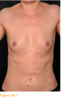
She has multiple naevi over her trunk and limbs, all of which have a similar appearance, with a slightly irregular border and varying shades of brown, tan and light red (Fig. 60.1).
Questions
• What is this syndrome?
• How would you manage this patient?
 Medicine
This patient was diagnosed with atypical (dysplastic) naevus syndrome. The patient has multiple atypical naevi, as do her family and a first-degree relative with a malignant melanoma.
Single atypical naevi are present in 5 per cent of the population. These naevi are usually larger than the rest of the patient’s moles and clinically have an asymmetrical shape, irregular border and outline. These moles tend to ‘stand-out from the crowd’. They have a greater variation in pigmentation than the rest of the patient’s moles. These atypical naevi tend to occur later in childhood than common, acquired melanocytic naevi. Histologically these moles show features of mild to severe architectural dysplasia. Atypical melanocytic naevi occur sporadically or as part of the familial atypical naevus syndrome (patients with this syndrome may have several hundred atypical naevi) and are potential precursors for malignant melanoma. To be diagnosed with the atypical naevus syndrome patients must have:
1. one or more first-degree or second-degree relatives with malignant melanoma;
2. a large number of naevi (often more than 50), some of which are atypical naevi clinically;
3. histologically dysplastic naevi.
Patients with atypical naevi have a slightly higher risk of developing melanoma than the general population, particularly if they have five or more. Patients with the atypical naevus syndrome have a far greater risk of developing melanoma than those with a small number of atypical moles.
Consequently, patients diagnosed with this syndrome should undergo life-long surveil-lance due to the potential risk of development of malignant melanoma. Patients are taught to examine their own skin and told what signs to look for, namely, change in size, shape or colour. Photographic and dermatoscopic records are helpful for monitoring these patients to detect early change. Any suspicious or changing atypical naevus should be removed by excision biopsy. Careful photoprotection and sun avoidance are essential.
KEY POINTS
• Atypical melanocytic naevi occur sporadically or as part of the familial atypical naevus syndrome.
• Patients with this syndrome have a far greater risk of developing melanoma.
• Patients diagnosed with this syndrome should undergo life-long surveillance.
need an explanation for this answer? contact us directly to get an explanation for this answer