


History
A 59-year-old man presents with a 7-week history of a rapidly growing lesion on the dorsum of his right hand. He had seen his GP who had referred him to the dermatology out-patient clinic. However, over the past week the patient had thought that the lesion was now getting smaller and had wondered about cancelling his appointment. His wife had insisted that he attend the clinic to put her mind at rest. He is otherwise well and denies any previous skin problems.
Examination
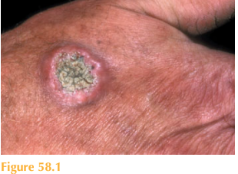
He has an erythematous, firm, dome-shaped nodule measuring 1.3 cm in diameter with a central keratin plug (Fig. 58.1). Full skin examination reveals a somewhat sun-damaged skin with freckling and solar lentigos but nothing else of note.

Questions
• What is the most likely diagnosis?
• What is the usual course and prognosis of this lesion?
 Medicine
The diagnosis of keratoacanthoma should always be considered when a patient presents with a rapidly enlarging nodule with a central crater. These lesions grow rapidly over a few weeks and are occasionally painful. They typically appear on sun-exposed sites, such as the face and the dorsal surfaces of the hands. Clinically they appear as dome-shaped nodules with shouldering that has a central keratin plug. If the plug falls away a crater is left. Although a keratoacanthoma may mimic a squamous cell carcinoma (SCC) the latter would not spontaneously regress which can occur with keratoacanthomas over a period of weeks to months.
Keratoacanthomas are epidermal tumours, which may behave in a benign way and are therefore called ‘pseudo’ tumours. In some cases there is a preceding history of minor trauma at the affected site. Previous ultraviolet light exposure appears to be a risk factor, as does human papilloma virus infections. Histologically, they can be difficult to distin-guish from SCCs as they contain cytologically atypical keratinocytes. For this reason they can usually only be distinguished on clinical grounds.
SCC is a skin cancer that can metastasize early into the blood. Therefore, most derma-tologists would excise such nodules to be sure they have always fully eradicated any potential SCCs. It is not current clinical practice to wait for keratoacanthomas to regress: they are treated as if they were SCCs.
In the right clinical context, where the suspicion of keratoacanthoma is very high and the patient deemed unsuitable for a full excision, some keratoacanthomas may be removed by curettage and cautery (tumour curettage – performed aggressively using three passes) with satisfactory results. If keratoacanthomas involute spontaneously, then they usually leave a cosmetically disfiguring residual scar. Hypertrophic actinic keratoses may also be included in the differential diagnosis.
Finally, a rare genetic disease called Ferguson–Smith syndrome is an autosomal domi-nant condition where multiple keratoacanthomas develop over the face and extremities.
In Ferguson–Smith syndrome the keratoacanthomas do regress spontaneously but lead to significant scarring.
KEY POINTS
• Keratoacanthomas manifest as dome-shaped nodules with a central keratin plug.
• Histologically they are difficult to distinguish from SCCs.
• Keratoacanthomas may spontaneously regress.
need an explanation for this answer? contact us directly to get an explanation for this answer