History
A 59-year-old construction worker presents to the dermatology clinic with a 3-year his-tory of a slow-growing lesion on the left temple. The patient describes that the lesion had begun as a small ‘spot’, which had gradually enlarged and had more recently been crusting over and intermittently bleeding. There is no previous history of skin disease and no family history of similar problems. He has worked outdoors for 40 years and has travelled abroad to tropical climates twice a year.
Examination
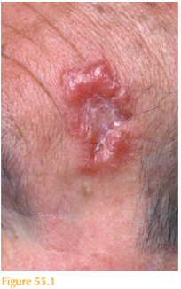
There is a nodular lesion 18 mm in diameter with a translucent edge and overlying tel-angiectasia with central crust (Fig. 55.1). Full skin examination does not reveal any other similar lesions, although he is noted as being tanned.

Questions
• What is the clinical diagnosis?
• What investigation should be performed?
• How should this patient be treated?
Clinically, this man has a basal cell carcinoma (BCC) or rodent ulcer. Malignant skin tumours are amongst the commonest of all cancers. BCCs are the most common of the skin cancer group and their incidence is continuing to rise. Most typically they are found on sun-exposed sites in lighter skinned, middle-aged to elderly individuals, although owing to changes in lifestyle and increasing travel such patients can present as early as in the third decade. There are different clinical and histological types. This patient has a nodular BCC, which is the most frequently seen and, like this lesion, has well-defined, rolled edges that have a pearly translucent appearance. On closer examination superficial blood vessels (telangiectasia) are often seen. As the BCC enlarges it can ulcerate, hence the patient’s description of recent intermittent bleeding and crusting. Eventually the ulceration can cause destruction of the underlying tissue. For example, around the nasal area the carti-lage can be destroyed and hence the name ‘rodent’ ulcer. This patient’s outdoor occupa-tion and recreational history of travel to areas of high intensity ultraviolet are important.
To confirm the diagnosis a skin biopsy was performed for histopathological confir-mation. Other subtypes include superficial BCCs, where the lesion is much flatter and erythematous, often with an overlying sur-face scale (Fig. 55.2). Morphoeic BCCs are translucent plaques with an ill-defined edge (Fig. 55.3).
Although BCCs have a low malignant poten-tial they are locally destructive and can cause significant morbidity, especially when occurring around facial structures such as the eyes and nose. Treatment is dependent on site, size and type of BCC. Treatment options include curettage and cautery, sur-gical excision, photodynamic therapy and radiotherapy. For those lesions in high-risk anatomical sites and for lesions that have clinically ill-defined margins, such as the morphoeic BCCs, then Mohs’ micrographic surgery is the treatment of choice, where the tissue is sectioned and examined to ensure that all the tumour has been excised before repairing the defect. Patients should undergo a full skin examination and be given future photoprotective advice.
KEY POINTS
• Basal cell carcinomas (BCCs) are the most common type of skin cancer.
• Subtypes of BCCs include nodular, superficial and morphoeic.
• Treatment options include curettage and cautery, surgical excision annd radiotherapy.
need an explanation for this answer? contact us directly to get an explanation for this answer