


History
A 3-year-old girl attends the paediatric dermatology clinic with her mother. She has a red lesion on her left ear, which her mother fears will lead to bullying in school. The lesion is entirely asymptomatic. Her parents first noticed the lesion shortly after birth and describe it as having the appearance of a red ‘stain’. Shortly before her six-week check it was beginning to enlarge and it grew until she was approximately four months old. Her mother feels it has been fairly static subsequently. The child is otherwise well; she was born at term and is fully vaccinated to date. There is no family history of birthmarks or other skin lesions.
Examination
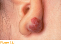
She is thriving with height and weight between the 50th and 75th centiles for her age. She has an elevated, dome-shaped, dusky red, rubbery, non-tender lesion on the dorsal aspect of the lobule of her left ear (Fig. 52.1). The overlying skin is showing some patchy pallor (or greying). She has no other skin lesions.
Questions
• What is this lesion?
• What is its natural history?
• What management options are available?
 Medicine
This lesion is an infantile haemangioma, also referred to as a capillary haemangioma, strawberry naevus or cavernous haemangioma. These are benign vascular neoplasms and are the most common tumours of infancy. Most infantile haemangiomas are medi-cally insignificant and the vast majority of lesions (80 per cent) are focal and solitary.
Occasionally, however, they can impinge on vital structures, ulcerate, bleed, become secondarily infected or painful; also they can cause high-output cardiac failure or signifi-cant structural abnormalities or disfigurement. Visual obstruction, airway obstruction or interference with feeding/defaecation are the most commonly encountered complications.
Haemangiomas can also occur in extracutaneous sites such as larynx, gastrointestinal tract and other abdominal viscera. Rarely, they may be associated with more extensive underlying congenital anomalies (e.g. PHACE syndrome, posterior fossa brain abnormali-ties, haemangiomas, arterial lesions, cardiac anomalies and eye abnormalities).
Infantile haemangiomas follow a characteristic course, with an early rapid-proliferation phase during the neonatal period or early infancy, followed by a slow gradual involution phase up to the age of approximately 10 years. Early features include blanching of the affected skin, fine telangiectases, or a red macule or papule. As they proliferate, depend-ing on their size and depth, their appearance may combine one or more features such as dome-shaped, lobulated, plaque-like, and tumoural. Most reach a maximum size of about 5 cm, but they can range from a pin-head to more than 20 cm in diameter. During the involution phase it is common for the haemangioma to shrink centrifugally from the centre; they become less red and gradually duskier (greying) before becoming softer and regaining flesh tones. The involution phase will be completed by 9 years of age in the overwhelming majority of patients, and for approximately 70 per cent of patients the haemangioma resolves completely. In the remainder there may be some residual perma-nent changes such as telangiectasia, superficial dilated vessels, stippled scarring, epider-mal atrophy, hypopigmentation and/or redundant skin with fibrofatty residua.
The majority of infantile haemangiomas, such as the case presented, do not require any medical or surgical intervention. Treatment is indicated to reduce morbidity and mortality and to prevent complications which may have an impact on growth and development.
In general the cosmetic aspect of haemangiomas is dealt with following the involution phase. Laser therapy is beneficial in treating ulcerated haemangiomas and thin superficial lesions in cosmetically sensitive sites. Surgical excision is exceptionally rare because of the potential intra-operative hazards and longer-term cosmetic results. Medical treatment with systemic or intra-lesional corticosteroids can be effective at slowing the growth and decreasing the size of proliferating haemangiomas. Propranolol is also emerging as a potentially more effective therapy during the proliferation phase, and has been in use for the management of severe or disfiguring haemangiomas since 2008. Duration of therapy varies from 2 to 10 months and there are currently no universally accepted criteria for initiation of therapy or therapeutic protocols. Propranolol, however, is very likely to make the use of agents such as interferon-and vincristine obsolete in the management of haemangiomas.
In the case presented it is essential to educate the parents about the natural history and prognosis of infantile haemangiomas, as well as the potential risks and benefits of differ-ent treatments. Emotional support and exchange of views are available through forums such as the Birthmark Society. However, there would be little indication for intervention at this stage.
KEY POINTS
• Infantile haemangiomas are the most common tumour of infancy.
• These benign endothelial neoplasms are typically absent at birth, they characteristically grow rapidly in infancy and gradually spontaneously involute over the first decade.
• The majority of haemangiomas require no treatment; however, if there are associated complications such as visual or airway obstruction, ulceration or bleeding, different treatments are available under close hospital supervision.
need an explanation for this answer? contact us directly to get an explanation for this answer