History
A 23-year-old woman attends the dermatology clinic complaining of the appearance of asymptomatic lesions on her legs. She has a background of type 1 diabetes mellitus, diagnosed at age 6 years. Her glycaemic control had been erratic when she first moved to university 4 years ago, however she now feels more in control of her glucose levels with careful titration of her insulin.
Examination
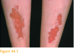
There are bilateral skin lesions affecting her anterior shins. The individual lesions are discrete and well demarcated with irregular borders. There is
atrophy of the dermis and epidermis resulting in a shiny (but ‘transparent’) surface with yellow colour and appar-ent arborizing telangiectasia (Fig.44.1). A full examination including fundoscopy was otherwise unremarkable. She has no skin lesions elsewhere.


Questions
• What are these lesions?
• What complications can be associated with them?
• How would you manage this patient?
This patient has a history of poor glycaemic control and evidence of diabetic nephropa-thy. Her current HbA1c value is acceptable but could be tighter. These skin lesions are called necrobiosis lipoidica (NL), which is associated with diabetes mellitus, particularly type 1. The frequency of diabetes mellitus amongst patients with NL ranges from 10 to 70 per cent in various studies, and the prevalence of NL amongst patients with diabetes mellitus ranges from 0.3 to 3.0 per cent. The association with poor glycaemic control is controversial. However, exercise and improved glycaemic control are reported to improve the lesions. The lesions initially present as red/brown papules and nodules which may resemble granuloma annulare or cutaneous sarcoid. They gradually flatten and become atrophic, taking on the characteristic appearance seen in Figure 44.1. NL has a predilec-tion for the anterior shins, but may also occur elsewhere on the lower legs and feet.
Although the clinical course may be indolent, NL lesions can be associated with refrac-tory ulceration. The clinical appearance and lack of spontaneous remission often prompts therapeutic intervention. Treatment of NL can be disappointing. Once established the hallmark of these lesions is atrophy and few therapeutic interventions improve their appearance. Early application of topical glucocorticoids may slow progression. Some authors advocate intralesional glucocorticoid injections, but these carry an associated risk of ulceration. Other treatment modalities described include phototherapy with psoralen–UVA and even aspirin. Ulcerated NL is managed with dressings and immunosuppressive therapy (ciclosporin, methotrexate and systemic glucocorticoids).
KEY POINTS
• Necrobiosis lipoidica (NL) is characterized by discrete atrophic shiny yellow patches with telangiectasis over the anterior shins.
• It can be associated with diabetes mellitus.
• The challenging aspects of NL include the cosmetic impact as well as lack of effective therapeutic modality.
• Ulceration is the most serious complication.
need an explanation for this answer? contact us directly to get an explanation for this answer