


History
A 32-year-old carpenter presents with a 3-month history of an itchy, and ‘stinging’ erup- tion. He had already been treated with a course of anti-scabies topical therapy and had used a moderately potent topical steroid in combination with antihistamine for 4 weeks with no benefit. He has no significant past medical history and is on no other medication. He comments that the eruption occurs in crops, with small fluid-filled blisters appearing in very itchy patches. He is one of five siblings, one of his sisters has type 1 diabetes mellitus and his mother is hypothyroid. On systems review he admits to fatigue and 4 kg unintentional weight loss associated with a change in stools over the past several months, which he attributes to stress at work.
Examination
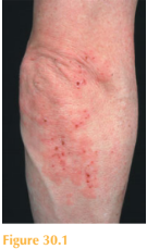
He has pale, freckled skin, and a symmetrical but patchy erup-tion, with erythematous and eczematous epidermal change studded with excoriated erosions and crusted papules, clustered particularly over his elbows (Fig. 30.1), buttocks and shoulders. He has no mucosal lesions and no evidence of scabies burrows. The remainder of his physical examination is normal.

All Answers
total answers (1)
 Medicine
The important clues to the diagnosis here are as follows: the distribution of the eruption; the history of pruritic vesicles; the positive family history of probable organ-specific autoimmune disease; his symptoms and blood test results suggestive of an underlying medical disorder. Although the clinical features in Fig. 30.1 are rather non-specific, the lack of response to the treatments already tried in combination with the clues before would all be suggestive of a diagnosis of dermatitis herpetiformis (DH). This is an autoim- mune blistering disease characterized by intensely itchy polymorphous vesicular lesions located over the extensor surfaces, back and scalp. It can be difficult to identify vesicles as they are often excoriated, but when the vesicles are present they can be clear, fluid- filled or pus-filled. A routine skin biopsy of peri-lesional skin will show papillary dermal neutrophilic microabcesses. Direct immunofluorescence shows immunoglobulin A (IgA) deposition within the papillary dermis. DH is a cutaneous manifestation of coeliac disease (gluten-sensitive enteropathy). Both conditions are immunological reactions to ingested gliadin (found in wheat, rye and barley). Serology to detect circulating anti-endomysial and anti-tissue transglutaminase IgA antibodies help to confirm the diagnosis, but a small bowel biopsy should also be performed. Occasionally the features of DH precede the pathological small bowel features of coeliac disease. A gluten-free diet (GFD) is the treatment of choice for both DH and coeliac disease; importantly it has a protective effect against small bowel lymphoma. Dapsone therapymay be required to gain control of the skin disease. However, after several months of compliance with a GFD this need is usually eliminated.
KEY POINTS
• Dermatitis herpetiformis (DH) is an autoimmune blistering disease and manifestation of gluten sensitive enteropathy.
• DH is associated with a personal or family history of other organ-specific autoimmune diseases.
• The characteristic eruption is composed of intensely pruritic vesicles clustering over extensor surfaces. However, secondary changes due to excoriation may predominate.
need an explanation for this answer? contact us directly to get an explanation for this answer