History
A 40-year-old woman is referred to the dermatology clinic with a history of erythematous skin lesions appearing on her face, neck and chest over the past six weeks. She complains of a burning and slight itching with the onset of new lesions that then settle over a few weeks leaving marked pigmentation on her skin. Intermittently she has developed new lesions in addition to the old lesions flaring. She had no previous history of skin prob-lems. Past medical history includes mild asthma, hayfever and hypertension. Her medi-cation includes becotide and salbutamol inhalers, simvastatin, atenolol and ramipril; the latter was commenced within the last 2 months to help control her hypertension.
Examination
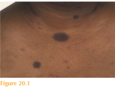
There are multiple annular hyperpigmented macular lesions on the skin over her face, neck and upper chest (Fig. 20.1). No new erythematous lesions or recently ‘reactivated’ lesions are apparent at the time of this clinic appointment.


Questions
• What is the diagnosis?
• What is the likely cause?
• How would you manage this patient?
This patient was diagnosed with a fixed drug eruption (FDE), which is an interesting phenomenon where exposure to a particular medication leads to annular erythematous skin lesion(s) which heal with pigmentation. Repeated exposure to the drug invariably causes reactivation of the old skin lesion(s) and may cause new ones to appear. Solitary skin lesions most commonly occur in FDE and preferentially occur on the genitalia and lips. This patient had the multiple FDE variant where lesions more frequently appear on the arms and trunk. There are reports of FDE lesions occurring on the genitals in patients who are allergic to their sexual partner’s medication. The morphology of the skin lesions in FDE is somewhat variable, however. Classically they are circular or oval, macular or bullous and heal with pigmentation. Some FDE erup-tions can be so inflammatory that central skin necrosis can result. The exact mechanism of FDE is not known. Our current understanding is that the medi-cation acts as a hapten which binds to keratinocytes within the epidermis. This triggers the release of pro-inflammatory cytokines which attract T-cells (CD4 and CD8) into the affected skin site. Reactivation of the skin lesions in response to re-exposure to the medi-cation activates memory T-cells at the identical skin site(s). Histological appearances of involved skin show the action is at the dermoepidermal junction. The onset of FDE usually starts within two weeks of commencing a new medication. In this case the most likely culprit is the ramipril owing to the temporal relationship between commencement of the drug and the onset of the skin lesions. The most common culprit drugs include antibiotics, anticonvulsants and analgesics. However, FDE has been reported as being associated with many other preparations including over-the-counter medications, supplements, minerals and even foods. The diagnosis of FDE is usually straightforward with a good history and classic clinical signs. If it is unclear which medication may be the culprit then an oral challenge can be useful. Histological appearances from lesional skin can help support the clinical diagnosis of FDE but is usually not diagnostic. Once the culprit medication has been identified this should be stopped. A potent topical steroid can be applied to any active lesions, which will help settle any symptoms and inflammation rapidly.
KEY POINTS
• FDE lesions on the skin are reactivated each time the culprit drug is ingested.
• Lesions are frequently solitary and have a predilection for the lips and genitals.
• Skin lesions are often annular with central blistering/necrosis that settles with pigmentation.
need an explanation for this answer? contact us directly to get an explanation for this answer