History
A 49-year-old man presents to the dermatology out-patient clinic with a 6-month his-tory of a gradually worsening rash on the dorsi of his hands. He had first noticed a few small blisters developing in the early spring and since then more have appeared. He is concerned about the scarring marks left on his hands. He has recently been made redun-dant and as a result has been drinking large quantities of alcohol. He has no history of previous skin problems or atopy. He is otherwise well and does not take any regular medication.
Examination
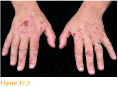
Over the dorsi of his hands are scattered tense vesicles and bullae with multiple erosions, scarring, milia and pigmentation (Fig. 17.1). Full skin examination reveals similar small scars on his face and minimal hypertrichosis. The rest of the skin is normal.

Questions
• What is the diagnosis?
• What is the underlying cause?
• How would you confirm the diagnosis?
This patient was not aware that he was photosensitive due to the insidious onset of his skin eruption over many months. Medical practitioners should suspect photosensitivity if skin disease affects exposed skin sites – classically the face, hands and anterior chest. On direct questioning, patients may report a worsening of their skin condition during the summer or following a holiday abroad. This patient was diagnosed with porphyria cutanea tarda (PCT) which usually presents between the 3rd to 5th decades. The condition is characterized by skin fragility leading to blister formation following minor trauma. Initially, skin changes include tense vesi-cles/bullae and erosions on a background of normal-looking skin. However, these heal to leave small atrophic scars and milia (inclusion cysts) as seen in this patient. Waxy-looking plaques and hypertrichosis may also be seen. Porphyrins are important in the formation of haemoglobin, myoglobin and cytochromes. The porphyrias are diseases in which specific enzyme deficiencies lead to the accumu-
lation of intermediate metabolites in the porphyrin biosynthesis pathway. Porphyrins absorb light in the 400–405 nm range (the lower range of visible light). This absorbed light is then transferred to cellular structures, causing damage to tissues. Deficiency of the enzyme uroporphyrinogen decarboxylase (UROD) is responsible for PCT, which may be inherited or acquired. Patients with acquired disease are deficient in UROD in their liver and therefore express the disease phenotypically when triggered by agents such as alcohol, oestrogen and hepatitis C. Familial PCT is inherited in an autosomal dominant fashion with a resultant defect in the synthesis of UROD. A skin biopsy reveals subepidermal bullae. Wood’s lamp examination of the urine shows orange red fluorescence. A porphyrin screen reveals raised uroporphyrin I in the urine and increased levels of isocoproporphyrins in the stool. Treatments include eliminating possible trigger factors such as alcohol. Patients should be protected from both ultraviolet and visible light. Management includes venesection to reduce iron stores or low-dose oral chloroquine/hydroxychloroquine.
KEY POINTS
• PCT occurs due to deficiency of the enzyme uroporphyrinogen decarboxylase.
• It may be inherited or acquired. Triggers include alcohol, oestrogen and hepatitis C.
• Skin changes include fragility, tense vesicles/bullae, erosions, atrophic scars and milia.
need an explanation for this answer? contact us directly to get an explanation for this answer