History
A 6-year-old boy is brought to the accident and emergency department by his parents with a 5-day history of worsening eczema associated with malaise and lethargy. In addi-tion to worsening pruritus and sleeplessness he complains of painful skin, particularly around his face, neck, chest and forearms. He quantifies the level of pain as 8 out of 10. His current flare is not responding to diligent application of his usual eczema treatments according to his ‘step-up’ management plan. The onset of his eczema was at the age of 4 months, and although moderately severe in infancy it has been reasonably controlled since starting primary school, with regular use of emollients and mild to moderately potent topical steroids. His background his-tory includes egg allergy (now partially outgrown in that he tolerates well cooked egg in cakes) and asthma, currently stable. He has never been admitted to hospital before. He is fully vaccinated to date and had chickenpox at the age of 4 years. His father suffers from hay-fever and experienced childhood eczema and asthma. He has one older sister (aged 14) who is well. His medication includes:
• Regular emollients both as leave-on preparations and soap substitute
• Topical tacrolimus 0.1% twice daily applied to affected areas for the management of flares
• Hydroxyzine 10 mg nocte during flares and salbutamol inhaler on a prn basis
Examination
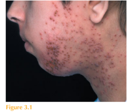
He looks unwell and is febrile at 38.5 °C. Systemic examination is normal except for widespread lymphadenopathy. There is no evidence of conjunctival erythema and his vision is normal. He has generalized moderate to severe eczema with erythema, dryness, excoriation and lichenification. He has a superimposed monomorphic eruption over his lower face, chest and forearms. The eruption is composed of multiple 23-mm monomor-phic ‘punched-out’ erythematous lesions in various stages of evolution (Fig. 3.1). Some of the lesions are vesicular, others pustular, some coalescing, most are eroded and covered with a golden exudate and others haemorrhagic crust.

Questions
• What are these lesions?
• How would you confirm the diagnosis?
• What complications can be associated with them?
• What is their management?
These are typical lesions of herpes simplex virus (HSV) infection complicating atopic eczema. This eruption is called eczema herpeticum, or less commonly Kaposi’s varicel-liform eruption. Diagnosis can be confirmed by viral swab of a blister or eroded area. Many tests can detect HSV within tissue or blister fluid. HSV can be inferred by positive staining or electron microscopy or specifically identified as types HSV-1 or HSV-2 by immunofluo-rescence, culture, or polymerase chain reaction. Bacteriology swab for microscopy and culture should also be undertaken. Significant morbidity is associated with eczema herpeticum. The main potential com-plications include superimposed bacterial infection (Staphylococcus or Streptococcus) with risk of systemic sepsis, ocular involvement (in particular, HSV keratitis) and, rarely, systemic HSV infection with risk of spread to the liver, the lungs, the brain, the gastroin-testinal tract and even the adrenal glands. In addition pain and discomfort associated with eczema herpeticum is significant. The management of widespread eczema herpeticum includes systemic treatment of HSV infection with aciclovir, identification and treatment of any superimposed bacterial infection or strategies to prevent superimposed infection, such as antibacterial washes and creams. Topical tacrolimus should be discontinued in this patient as this may exac-erbate the cutaneous spread of HSV. These cases are usually managed as in-patients, initially with intravenous aciclovir – as oral preparations can be poorly absorbed. Ophthamological review should be sought in cases of diffuse facial herpes simplex infec-tion or where conjunctival/corneal involvement is suspected. In a minority of cases recurrences can occur. Rapid treatment of incipient lesions with topical aciclovir may help prevent disseminated eczema herpeticum.
KEY POINTS
• Herpes simplex infection in patients with eczema can lead to widespread lesions and an associated risk of superimposed bacterial infection and sepsis.
• It is important to consider and exclude the rare associated complication of herpes keratitis.
• In-patient management with systemic anti-viral therapy, topical antiseptic measures, pain relief, and where indicated antibiotic therapy, is required.
need an explanation for this answer? contact us directly to get an explanation for this answer