


FEVER AND BREATHLESSNESS
History
Niall is a 3-year-old boy from a travelling family referred to the paediatric day unit by the out-of-hours GP service. He was seen 4 days ago with a cough and fever and diagnosed with a viral upper respiratory tract infection (URTI). The following day he returned and was commenced on oral antibiotics. He is now complaining of tummy ache and has vomited once. The GP is worried that he is becoming dehydrated and may have a urinary tract infection or intra-abdominal pathology. He has not been immunized but there is no other medical history of note.
Examination Niall is miserable, flushed, toxic and febrile (38.8C) with a capillary refill time of 2 s. His pulse is 140 beats/min, his oxygen saturation is 91 per cent in air and his blood pressure is 85/60 mmHg. He seems to be in pain, especially when he coughs, and his respiratory rate is 48 breaths/min with nasal flaring. There is dullness to percussion in the right lower zone posteriorly with decreased breath sounds and bronchial breathing. He seems reluctant to have his abdomen examined but bowel sounds are normal.
INVESTIGATIONS
Haemoglobin 11.8 g/dL 11.5–15.5 g/dL White cell count 25.4 109/L 6–17.5 109/L
Neutrophils 20.8 109/L 3–5.8 109/L Platelets 467 109/L 150–400 109/L
Sodium 126 mmol/L 138–146 mmol/L
Potassium 3.5 mmol/L 3.5–5.0 mmol/L Urea 2.5 mmol/L 1.8–6.4 mmol/L
Creatinine 48 µmol/L 27–62 µmol/L
Glucose 4.5 mmol/L 3.3–5.5 mmol/L
C-reactive protein 387 mg/L 6 mg/L Chest X-ray – see Figure 7.1
Questions
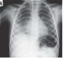
• What are the chest X-ray findings?
• What is the most likely causative organism?
• What complication may have arisen and how would you confirm it?
• List the steps in management.
 Medicine
The chest X-ray shows loss of the right hemidiaphragm, right lower zone consolidation and a normal right heart border – the characteristic features of right lower lobe pneumonia. Lower lobe pneumonia should always be on the list of differential diagnoses for abdominal pain in children. Young children rarely localize pain but observation should establish whether they have pleuritic pain – they may have shallow breathing or may simply sit very still, hardly moving the affected side. The most likely causative organism is Streptococcus pneumoniae. However, a diagnosis is rarely made from sputum analysis because children tend to swallow their sputum. Blood cultures may be positive. Children in the UK are immunized against Streptococcus pneumoniae but not all strains are covered, there is emerging antibiotic resistance and also ‘failed’ immunizations. Any immunized child with positive cultures should be investigated for an underlying immune problem, including an absent spleen. Niall has hyponatraemia. There are no pointers to excess sodium loss (e.g. diarrhoea or significant vomiting) and hence the most likely cause is the syndrome of inappropriate antidiur etic hormone secretion (SIADH), a known association with pneumonia. The hyponatraemia is dilutional. First, the result should be confirmed – taking blood from children can be difficult and unexpected results should be repeated. At the same time, urine should be sent for osmolality and sodium. His serum osmolality is calculated as follows: 2 ([Na] [K]) [urea] [glucose] 266 mosmol/kg (normal 278–305) Normally a fall in serum osmolality would suppress antidiuretic hormone secretion to allow excretion of excess water as dilute urine. In SIADH, urine osmolality is inappropriately high (320 mosmol/kg) and urine sodium is usually 40 mmol/L (unlike hypovolaemic states where it is 20 mmol/L).
Steps in management
• Oxygen to maintain saturation at 92 per cent
• Adequate pain relief for pleuritic pain
• Intravenous antibiotics according to local guidelines, e.g. co-amoxiclav
• Initial fluid restriction to two-thirds maintenance to help correct the hyponatraemia. Fluid restrict even if no hyponatraemia, as SIADH may still develop
• Fluid balance, regular urea and electrolytes – adjust fluids accordingly. Weigh twice daily
• Physiotherapy, e.g. bubble blowing. Encourage mobility
• Monitor for development of a pleural effusion. If the chest X-ray is suspicious, an ultrasound will be diagnostic. If present, a longer course of antibiotics is recommended to prevent empyema (a purulent pleural effusion). A chest drain may be necessary if there is worsening respiratory distress, mediastinal shift on the chest X-ray, a large effusion or failure to respond to adequate antibiotics
• Ensure adequate nutrition – children have often been anorectic for several days. Low threshold for supplementary feeds probably via nasogastric tube
• Organise immunization programme before discharge
• Arrange a follow-up chest X-ray in 6–8 weeks for those with lobar collapse and/ or an effusion. If still abnormal, consider an inhaled foreign body.
KEY POINTS
• A lower lobe pneumonia should always be considered in the differential diagnosis of an acute abdomen in children.
• ‘Vaccine failures’ should be investigated for an underlying immune problem.
need an explanation for this answer? contact us directly to get an explanation for this answer