


0
Figure 12.11 shows the visual field of a normal left eye as plotted by perimetry. When the eye is focused on point Y, an object at point:
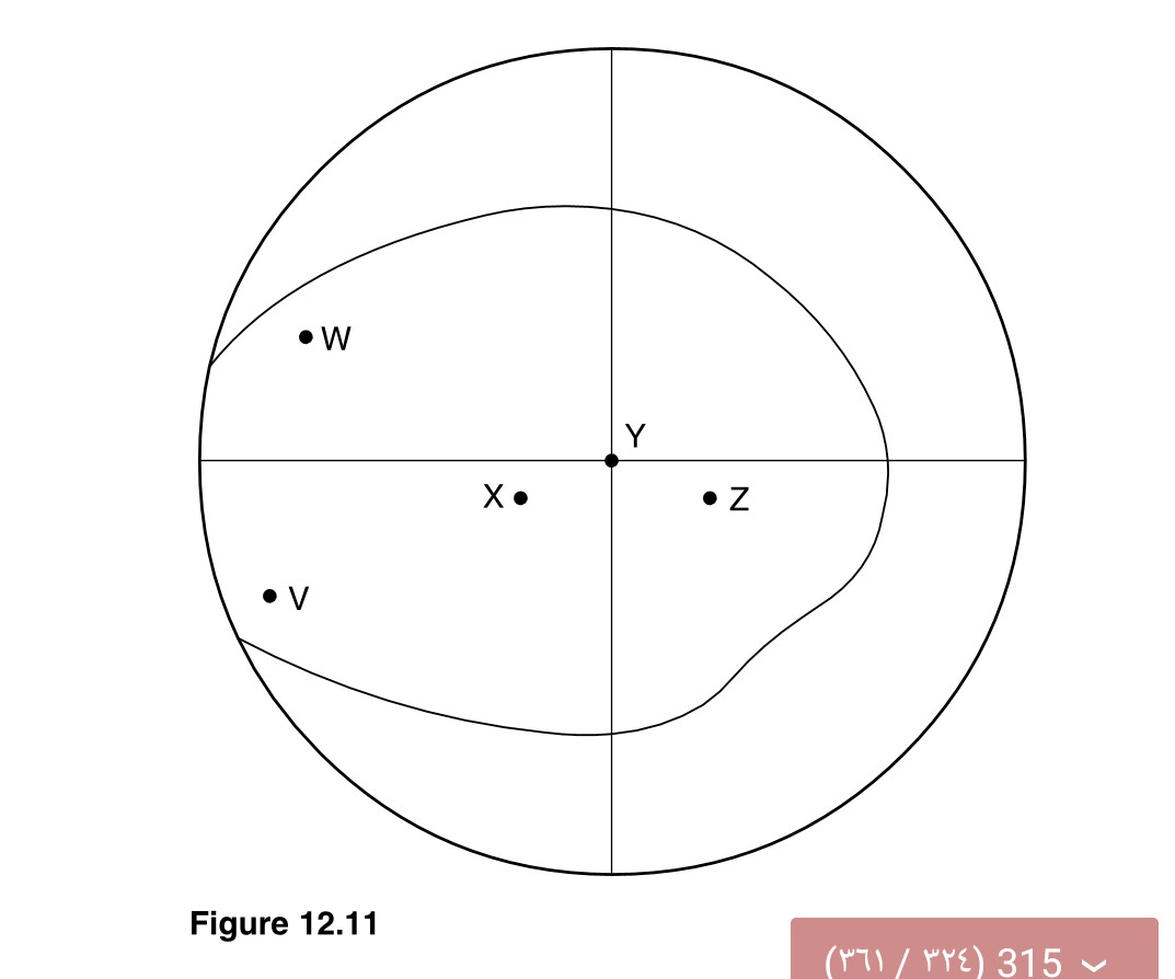
- W is detected in the lower nasal quadrant of the left retina.
- Y is detected in the region of the fovea of the macula.
- . Z rather than at point X may be invisible.
- W is appreciated as a result of impulses transmitted in the left rather than the right optic tract.
- V is seen in monocular vision.
 Medicine
a. True The image is inverted and reversed with respect to the object.
b. True The point focused upon is detected at the macula where visual acuity is greatest.
c. False The reverse is the case; the optic disc is medial to the fovea, hence the blind spot is in the temporal (lateral) part of the field of vision.
d. False Impulses from the temporal region of the field of vision cross the midline at the optic chiasma.
e. True The visual fields of the two eyes do not overlap for this point.
need an explanation for this answer? contact us directly to get an explanation for this answer